X-rays: Looking inside our pets!
(Once you've read all this, click here to go help Dr. Chuck read some radiographs and save the day!)
X-rays are used to look inside our pets. First some clarification: An X-ray is a type of radiation (think of it as a special type of light, like a sun beam or a laser beam). A radiograph is a picture we take using X-rays.
To take a radiograph, we put our pet on a table, and then send X-ray beams through our pet and onto a piece of film. Our pet’s body blocks some of the X-rays, just like your hand can block light and create a shadow. Unlike normal light, however, X-rays can pass through parts of the body. X-rays can pass through air, but do not pass through bone, so the bones show up like shadows in the radiograph. X-rays can partly pass through water, fat, and tissue (the squishy parts of our bodies), so these make partial shadows in the radiograph. To make matters more confusing, because of how we develop radiographs, the 'shadow' in a radiograph is white, while the rest of the picture is black!
A regular shadow is dark!
An X-ray 'shadow' is white! The bones block the X-rays more than the rest of the foot, so the bones are white and the rest of the foot is gray.
So how do we use this information to look at radiographs? When we look at the radiograph, anything mostly white is bone or mineral (very bright white is metal, like if your dog ate his name tag!). Next brightest is fluid and soft tissue (like your bladder when you have to pee, or your kidneys). Slightly darker than water is fat, which is found all around the body. Darkest is air, which we see in the lungs, or in an upset stomach!
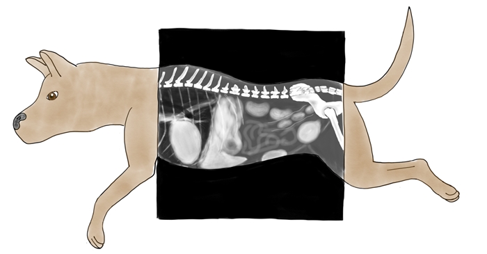
Here we can see the white, black, and shades of gray that come out in a radiograph! Notice how the area around the outside of the dog is black; this is because air is black in a radiograph! Anywhere the dog is, the radiograph is grey or white, because the dog made a 'shadow' in the X-ray beam.
A radiograph is also a great way to find an egg!
When a veterinarian looks at a radiograph, we say they are reading the radiograph. A picture is worth a thousand words! A veterinarian can look for many things in a radiograph; a broken bone, a swallowed ball, a full bladder (better let your dog go pee!), and much more!
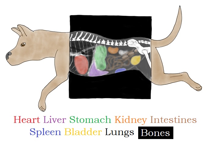
It takes a very long time to learn how to read a radiograph; they don't even come in color! But to make it easier, here we have labeled the major organs in our patient so you can see where they are in the body.
Red: This is the heart! The heart moves blood around the body.
Green: This is the stomach! This is where food goes when you eat!
Purple: This is the liver! The liver helps filter toxins out of the blood.
Orange: This is a kidney! Kidneys produce urine (pee!). We only colored in one kidney; the other kidney is right next to it.
Brown: These are the intestines! Can you spot the future poop?
Yellow: This is the bladder! The bladder holds pee until we can find a bathroom! Sometimes we find bright white bladder stones here.
Blue: This is the spleen! The spleen plays a big role in keeping our red and white blood cells in order, and stores extra red blood cells.
White: These are the bones! You can see the spine along the top of the picture, and part of the hips and femur (the big leg bone) on the far right. The ribs on the left of the picture are very thin, so they are not as bright as the other bones.
Black: The darkest part of the radiograph inside the dog is on the far left, where the lungs sit around the heart! Lungs are full of air, which makes them black.
Think you've got it all figured out?


